The Brain Belongs to What Division of the Nervous System
The Nervous System and Nervous Tissue
Bones Structure and Function of the Nervous Arrangement
Learning Objectives
Past the end of this section, yous will be able to:
- Identify the anatomical and functional divisions of the nervous system
- Relate the functional and structural differences between gray matter and white affair structures of the nervous system to the structure of neurons
- List the bones functions of the nervous system
The pic you have in your mind of the nervous organisation probably includes the brain, the nervous tissue contained within the cranium, and the spinal string, the extension of nervous tissue within the vertebral column. That suggests it is made of 2 organs—and you may not even think of the spinal cord as an organ—but the nervous system is a very circuitous construction. Within the brain, many different and separate regions are responsible for many different and separate functions. It is every bit if the nervous arrangement is composed of many organs that all wait similar and can simply be differentiated using tools such every bit the microscope or electrophysiology. In comparison, it is like shooting fish in a barrel to see that the stomach is different than the esophagus or the liver, so yous can imagine the digestive system as a collection of specific organs.
The Key and Peripheral Nervous Systems
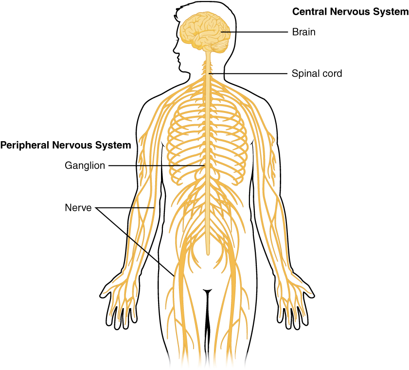
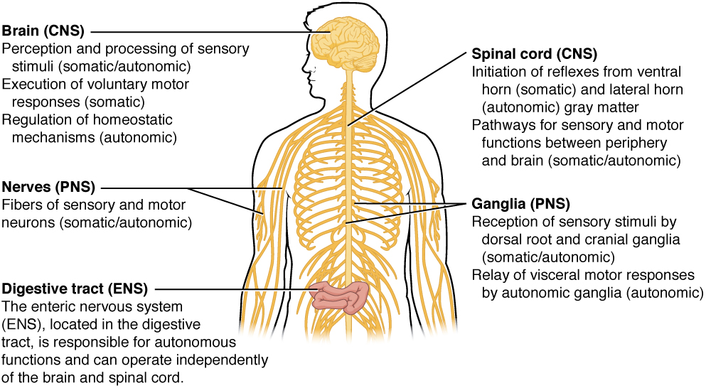
The nervous system can be divided into ii major regions: the central and peripheral nervous systems. The primal nervous system (CNS) is the brain and spinal cord, and the peripheral nervous system (PNS) is everything else ((Figure)). The brain is contained within the cranial cavity of the skull, and the spinal cord is contained inside the vertebral crenel of the vertebral cavalcade. It is a bit of an oversimplification to say that the CNS is what is inside these two cavities and the peripheral nervous system is outside of them, but that is 1 way to start to think about it. In authenticity, in that location are some elements of the peripheral nervous arrangement that are within the cranial or vertebral cavities. The peripheral nervous organisation is so named because it is on the periphery—meaning across the brain and spinal string. Depending on dissimilar aspects of the nervous system, the dividing line between central and peripheral is not necessarily universal.
Central and Peripheral Nervous System
The structures of the PNS are referred to every bit ganglia and nerves, which can be seen as singled-out structures. The equivalent structures in the CNS are not obvious from this overall perspective and are best examined in prepared tissue under the microscope.
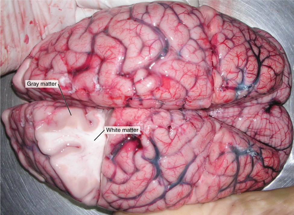
Nervous tissue, present in both the CNS and PNS, contains two basic types of cells: neurons and glial cells. A glial cell is ane of a diversity of cells that provide a framework of tissue that supports the neurons and their activities. The neuron is the more than functionally important of the two, in terms of the chatty role of the nervous organisation. To describe the functional divisions of the nervous system, it is of import to understand the structure of a neuron. Neurons are cells and therefore have a soma, or cell torso, merely they also have extensions of the cell; each extension is generally referred to equally a process. At that place is one important process that every neuron has called an axon, which is the fiber that connects a neuron with its target. Another type of process that branches off from the soma is the dendrite. Dendrites are responsible for receiving near of the input from other neurons. Looking at nervous tissue, in that location are regions that predominantly contain cell bodies and regions that are largely composed of merely axons. These ii regions within nervous system structures are oft referred to as greyness thing (the regions with many prison cell bodies and dendrites) or white affair (the regions with many axons). (Figure) demonstrates the appearance of these regions in the brain and spinal cord. The colors ascribed to these regions are what would be seen in "fresh," or unstained, nervous tissue. Gray matter is not necessarily gray. Information technology can be pinkish because of blood content, or even slightly tan, depending on how long the tissue has been preserved. But white matter is white considering axons are insulated by a lipid-rich substance chosen myelin. Lipids can appear equally white ("fatty") material, much like the fat on a raw piece of chicken or beef. Actually, grayness affair may have that color ascribed to it because adjacent to the white matter, information technology is just darker—hence, gray.
The distinction between gray affair and white matter is most oft practical to fundamental nervous tissue, which has large regions that can be seen with the unaided eye. When looking at peripheral structures, frequently a microscope is used and the tissue is stained with artificial colors. That is not to say that fundamental nervous tissue cannot be stained and viewed under a microscope, but unstained tissue is most likely from the CNS—for example, a frontal section of the brain or cross section of the spinal string.
Grayness Affair and White Affair
A brain removed during an autopsy, with a partial department removed, shows white affair surrounded by gray matter. Grayness affair makes up the outer cortex of the brain. (credit: modification of piece of work past "Suseno"/Wikimedia Commons)
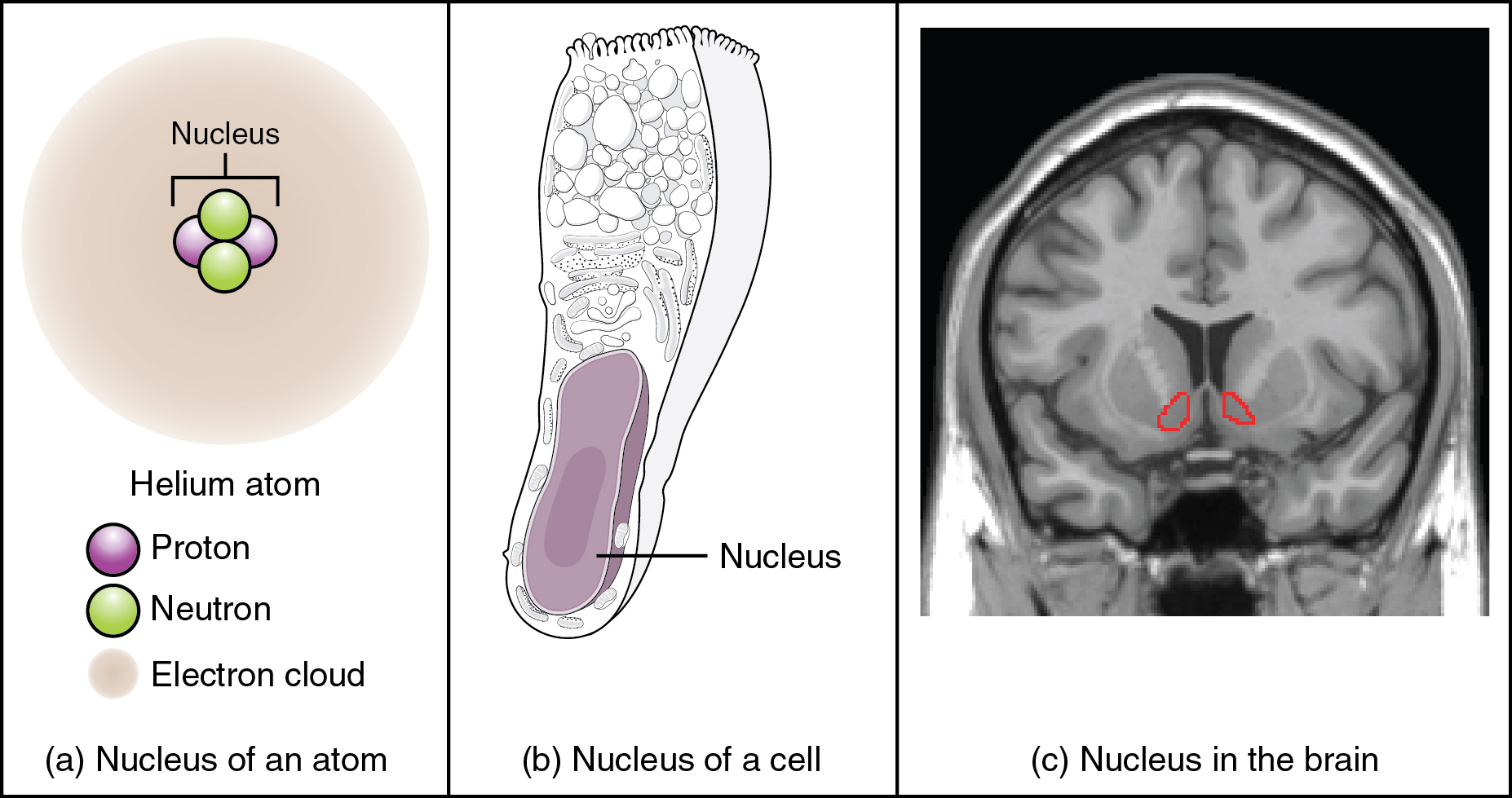
Regardless of the advent of stained or unstained tissue, the cell bodies of neurons or axons tin can exist located in discrete anatomical structures that need to be named. Those names are specific to whether the structure is key or peripheral. A localized drove of neuron jail cell bodies in the CNS is referred to as a nucleus. In the PNS, a cluster of neuron cell bodies is referred to as a ganglion. (Figure) indicates how the term nucleus has a few different meanings within anatomy and physiology. It is the centre of an atom, where protons and neutrons are found; information technology is the center of a prison cell, where the DNA is plant; and it is a center of some function in the CNS. There is also a potentially confusing use of the word ganglion (plural = ganglia) that has a historical caption. In the central nervous system, there is a grouping of nuclei that are continued together and were one time called the basal ganglia before "ganglion" became accepted as a description for a peripheral structure. Some sources refer to this group of nuclei as the "basal nuclei" to avoid confusion.
What Is a Nucleus?
(a) The nucleus of an cantlet contains its protons and neutrons. (b) The nucleus of a cell is the organelle that contains DNA. (c) A nucleus in the CNS is a localized heart of function with the cell bodies of several neurons, shown hither circled in red. (credit c: "Was a bee"/Wikimedia Eatables)
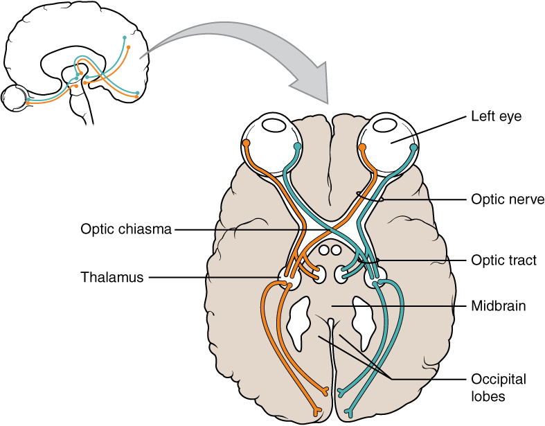
Terminology applied to bundles of axons likewise differs depending on location. A bundle of axons, or fibers, establish in the CNS is called a tract whereas the same matter in the PNS would be called a nerve. There is an important point to make well-nigh these terms, which is that they can both be used to refer to the same package of axons. When those axons are in the PNS, the term is nervus, but if they are CNS, the term is tract. The most obvious instance of this is the axons that projection from the retina into the brain. Those axons are chosen the optic nerve equally they leave the eye, simply when they are within the cranium, they are referred to as the optic tract. There is a specific place where the name changes, which is the optic chiasm, simply they are all the same the same axons ((Figure)). A similar situation outside of science can be described for some roads. Imagine a road chosen "Broad Street" in a boondocks called "Anyville." The road leaves Anyville and goes to the next town over, called "Hometown." When the road crosses the line between the two towns and is in Hometown, its name changes to "Main Street." That is the idea behind the naming of the retinal axons. In the PNS, they are called the optic nerve, and in the CNS, they are the optic tract. (Effigy) helps to clarify which of these terms apply to the central or peripheral nervous systems.
Optic Nerve Versus Optic Tract
This drawing of the connections of the heart to the brain shows the optic nerve extending from the eye to the chiasm, where the structure continues equally the optic tract. The same axons extend from the eye to the brain through these two bundles of fibers, just the chiasm represents the border betwixt peripheral and central.
In 2003, the Nobel Prize in Physiology or Medicine was awarded to Paul C. Lauterbur and Sir Peter Mansfield for discoveries related to magnetic resonance imaging (MRI). This is a tool to see the structures of the body (non just the nervous system) that depends on magnetic fields associated with certain atomic nuclei. The utility of this technique in the nervous organization is that fat tissue and water appear as unlike shades betwixt black and white. Because white matter is fatty (from myelin) and gray thing is not, they tin can exist easily distinguished in MRI images. Visit the Nobel Prize web site to play an interactive game that demonstrates the employ of this technology and compares it with other types of imaging technologies. As well, the results from an MRI session are compared with images obtained from Ten-ray or computed tomography. How do the imaging techniques shown in this game indicate the separation of white and gray matter compared with the freshly dissected tissue shown earlier?
| Structures of the CNS and PNS | ||
|---|---|---|
| CNS | PNS | |
| Grouping of Neuron Cell Bodies (i.e., grey matter) | Nucleus | Ganglion |
| Package of Axons (i.east., white matter) | Tract | Nerve |
Functional Divisions of the Nervous System
The nervous arrangement tin can also be divided on the basis of its functions, but anatomical divisions and functional divisions are different. The CNS and the PNS both contribute to the aforementioned functions, but those functions can be attributed to unlike regions of the brain (such equally the cerebral cortex or the hypothalamus) or to different ganglia in the periphery. The problem with trying to fit functional differences into anatomical divisions is that sometimes the same construction can be part of several functions. For example, the optic nerve carries signals from the retina that are either used for the conscious perception of visual stimuli, which takes place in the cerebral cortex, or for the reflexive responses of smooth muscle tissue that are processed through the hypothalamus.
In that location are ii ways to consider how the nervous system is divided functionally. First, the basic functions of the nervous organisation are sensation, integration, and response. Secondly, control of the body tin be somatic or autonomic—divisions that are largely defined by the structures that are involved in the response. There is also a region of the peripheral nervous system that is called the enteric nervous organization that is responsible for a specific set of the functions within the realm of autonomic control related to gastrointestinal functions.
Bones Functions
The nervous organisation is involved in receiving information about the surroundings around u.s.a. (awareness) and generating responses to that information (motor responses). The nervous system can be divided into regions that are responsible for sensation (sensory functions) and for the response (motor functions). But at that place is a third role that needs to be included. Sensory input needs to exist integrated with other sensations, likewise every bit with memories, emotional land, or learning (noesis). Some regions of the nervous system are termed integration or association areas. The procedure of integration combines sensory perceptions and higher cognitive functions such equally memories, learning, and emotion to produce a response.
Sensation. The showtime major function of the nervous organisation is sensation—receiving information about the environment to proceeds input about what is happening outside the body (or, sometimes, inside the torso). The sensory functions of the nervous system annals the presence of a change from homeostasis or a particular event in the environment, known as a stimulus. The senses we think of most are the "big 5": taste, odour, bear upon, sight, and hearing. The stimuli for taste and odor are both chemical substances (molecules, compounds, ions, etc.), touch is physical or mechanical stimuli that collaborate with the skin, sight is calorie-free stimuli, and hearing is the perception of sound, which is a physical stimulus similar to some aspects of touch. There are really more senses than but those, but that list represents the major senses. Those v are all senses that receive stimuli from the outside earth, and of which in that location is conscious perception. Additional sensory stimuli might be from the internal surroundings (inside the body), such as the stretch of an organ wall or the concentration of sure ions in the blood.
Response. The nervous arrangement produces a response on the footing of the stimuli perceived by sensory structures. An obvious response would be the move of muscles, such as withdrawing a paw from a hot stove, but at that place are broader uses of the term. The nervous system tin crusade the wrinkle of all three types of muscle tissue. For example, skeletal muscle contracts to motility the skeleton, cardiac muscle is influenced every bit heart charge per unit increases during practice, and polish muscle contracts as the digestive organization moves food forth the digestive tract. Responses too include the neural control of glands in the body every bit well, such as the production and secretion of sweat past the eccrine and merocrine sweat glands found in the skin to lower torso temperature.
Responses can be divided into those that are voluntary or conscious (contraction of skeletal musculus) and those that are involuntary (contraction of shine muscles, regulation of cardiac musculus, activation of glands). Voluntary responses are governed by the somatic nervous system and involuntary responses are governed past the autonomic nervous system, which are discussed in the next section.
Integration. Stimuli that are received by sensory structures are communicated to the nervous system where that information is processed. This is called integration. Stimuli are compared with, or integrated with, other stimuli, memories of previous stimuli, or the state of a person at a particular fourth dimension. This leads to the specific response that will be generated. Seeing a baseball pitched to a batter will not automatically cause the batter to swing. The trajectory of the ball and its speed volition demand to be considered. Possibly the count is iii balls and i strike, and the concoction wants to let this pitch become past in the promise of getting a walk to first base of operations. Or maybe the concoction's team is so far alee, it would be fun to just swing abroad.
Controlling the Body
The nervous arrangement tin be divided into two parts mostly on the basis of a functional deviation in responses. The somatic nervous organization (SNS) is responsible for conscious perception and voluntary motor responses. Voluntary motor response ways the contraction of skeletal muscle, but those contractions are not e'er voluntary in the sense that you have to desire to perform them. Some somatic motor responses are reflexes, and often happen without a witting decision to perform them. If your friend jumps out from behind a corner and yells "Boo!" y'all will be startled and you might scream or bound back. You didn't determine to do that, and yous may non have wanted to give your friend a reason to express mirth at your expense, but it is a reflex involving skeletal musculus contractions. Other motor responses get automated (in other words, unconscious) every bit a person learns motor skills (referred to as "addiction learning" or "procedural memory").
The autonomic nervous system (ANS) is responsible for involuntary control of the body, usually for the sake of homeostasis (regulation of the internal environment). Sensory input for autonomic functions tin can be from sensory structures tuned to external or internal environmental stimuli. The motor output extends to smooth and cardiac muscle as well as glandular tissue. The role of the autonomic system is to regulate the organ systems of the body, which normally ways to control homeostasis. Sweat glands, for example, are controlled by the autonomic system. When you lot are hot, sweating helps cool your body downwards. That is a homeostatic mechanism. But when you are nervous, you lot might kickoff sweating also. That is non homeostatic, it is the physiological response to an emotional state.
At that place is some other sectionalisation of the nervous system that describes functional responses. The enteric nervous organisation (ENS) is responsible for decision-making the smoothen muscle and glandular tissue in your digestive system. It is a big function of the PNS, and is not dependent on the CNS. It is sometimes valid, however, to consider the enteric organization to be a part of the autonomic arrangement because the neural structures that brand up the enteric system are a component of the autonomic output that regulates digestion. There are some differences between the 2, but for our purposes here there will exist a good bit of overlap. See (Figure) for examples of where these divisions of the nervous system can be found.
Somatic, Autonomic, and Enteric Structures of the Nervous Organisation
Somatic structures include the spinal fretfulness, both motor and sensory fibers, equally well every bit the sensory ganglia (posterior root ganglia and cranial nervus ganglia). Autonomic structures are plant in the nerves besides, simply include the sympathetic and parasympathetic ganglia. The enteric nervous system includes the nervous tissue inside the organs of the digestive tract.
Visit this site to read about a adult female that notices that her daughter is having trouble walking up the stairs. This leads to the discovery of a hereditary condition that affects the brain and spinal string. The electromyography and MRI tests indicated deficiencies in the spinal cord and cerebellum, both of which are responsible for decision-making coordinated movements. To what functional division of the nervous organization would these structures vest?
Everyday Connection
How Much of Your Brain Practice You Use? Accept you ever heard the claim that humans just employ 10 percent of their brains? Maybe you have seen an advertisement on a website proverb that there is a secret to unlocking the full potential of your mind—every bit if there were 90 percentage of your brain sitting idle, merely waiting for you to apply information technology. If you see an advert like that, don't click. Information technology isn't truthful.
An easy way to run into how much of the brain a person uses is to have measurements of brain activity while performing a task. An example of this kind of measurement is functional magnetic resonance imaging (fMRI), which generates a map of the most active areas and tin be generated and presented in three dimensions ((Effigy)). This procedure is dissimilar from the standard MRI technique because information technology is measuring changes in the tissue in time with an experimental condition or event.
fMRI
This fMRI shows activation of the visual cortex in response to visual stimuli. (credit: "Superborsuk"/Wikimedia Commons)
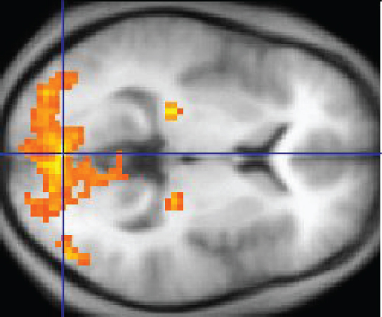
The underlying assumption is that active nervous tissue will accept greater blood menstruum. By having the subject perform a visual chore, action all over the brain tin be measured. Consider this possible experiment: the discipline is told to look at a screen with a blackness dot in the centre (a fixation bespeak). A photograph of a face is projected on the screen away from the heart. The subject has to await at the photograph and decipher what it is. The subject has been instructed to button a push button if the photograph is of someone they recognize. The photograph might be of a glory, so the bailiwick would press the button, or it might exist of a random person unknown to the subject area, so the subject area would not press the button.
In this task, visual sensory areas would be agile, integrating areas would be active, motor areas responsible for moving the optics would exist active, and motor areas for pressing the button with a finger would exist agile. Those areas are distributed all around the brain and the fMRI images would show activity in more than just x percent of the brain (some evidence suggests that well-nigh fourscore percent of the brain is using energy—based on blood flow to the tissue—during well-defined tasks similar to the 1 suggested above). This task does not fifty-fifty include all of the functions the brain performs. There is no language response, the body is mostly lying still in the MRI machine, and it does not consider the autonomic functions that would exist ongoing in the groundwork.
Chapter Review
The nervous system tin be separated into divisions on the basis of anatomy and physiology. The anatomical divisions are the central and peripheral nervous systems. The CNS is the brain and spinal cord. The PNS is everything else. Functionally, the nervous organisation can be divided into those regions that are responsible for awareness, those that are responsible for integration, and those that are responsible for generating responses. All of these functional areas are found in both the central and peripheral anatomy.
Considering the anatomical regions of the nervous system, in that location are specific names for the structures within each division. A localized collection of neuron cell bodies is referred to equally a nucleus in the CNS and every bit a ganglion in the PNS. A parcel of axons is referred to as a tract in the CNS and equally a nerve in the PNS. Whereas nuclei and ganglia are specifically in the central or peripheral divisions, axons can cross the boundary between the two. A unmarried axon can be part of a nervus and a tract. The proper noun for that specific structure depends on its location.
Nervous tissue can also exist described every bit gray thing and white matter on the basis of its appearance in unstained tissue. These descriptions are more oft used in the CNS. Grey matter is where nuclei are found and white matter is where tracts are found. In the PNS, ganglia are basically gray matter and nerves are white matter.
The nervous system can also be divided on the ground of how information technology controls the body. The somatic nervous organization (SNS) is responsible for functions that result in moving skeletal muscles. Whatsoever sensory or integrative functions that outcome in the motion of skeletal muscle would be considered somatic. The autonomic nervous system (ANS) is responsible for functions that affect cardiac or smooth musculus tissue, or that crusade glands to produce their secretions. Autonomic functions are distributed between key and peripheral regions of the nervous system. The sensations that lead to autonomic functions tin be the aforementioned sensations that are part of initiating somatic responses. Somatic and autonomic integrative functions may overlap as well.
A special partitioning of the nervous arrangement is the enteric nervous organization, which is responsible for decision-making the digestive organs. Parts of the autonomic nervous system overlap with the enteric nervous organisation. The enteric nervous system is exclusively found in the periphery considering it is the nervous tissue in the organs of the digestive arrangement.
Interactive Link Questions
In 2003, the Nobel Prize in Physiology or Medicine was awarded to Paul C. Lauterbur and Sir Peter Mansfield for discoveries related to magnetic resonance imaging (MRI). This is a tool to see the structures of the body (not just the nervous organization) that depends on magnetic fields associated with certain diminutive nuclei. The utility of this technique in the nervous system is that fat tissue and water announced as unlike shades between blackness and white. Because white matter is fatty (from myelin) and gray matter is non, they tin be easily distinguished in MRI images. Visit the Nobel Prize website to play an interactive game that demonstrates the use of this engineering and compares it with other types of imaging technologies. Also, the results from an MRI session are compared with images obtained from x-ray or computed tomography. How do the imaging techniques shown in this game point the separation of white and gray matter compared with the freshly dissected tissue shown earlier?
MRI uses the relative amount of water in tissue to distinguish unlike areas, and so gray and white matter in the nervous arrangement tin be seen conspicuously in these images.
Visit this site to read nearly a adult female that notices that her girl is having trouble walking upward the stairs. This leads to the discovery of a hereditary condition that affects the brain and spinal cord. The electromyography and MRI tests indicated deficiencies in the spinal cord and cerebellum, both of which are responsible for controlling coordinated movements. To what functional division of the nervous organisation would these structures belong?
They are part of the somatic nervous system, which is responsible for voluntary movements such as walking or climbing the stairs.
Review Questions
Which of the following cavities contains a component of the central nervous system?
- intestinal
- pelvic
- cranial
- thoracic
Which structure predominates in the white thing of the brain?
- myelinated axons
- neuronal cell bodies
- ganglia of the parasympathetic fretfulness
- bundles of dendrites from the enteric nervous organization
Which office of a neuron transmits an electrical signal to a target cell?
- dendrites
- soma
- cell body
- axon
Which term describes a bundle of axons in the peripheral nervous system?
- nucleus
- ganglion
- tract
- nervus
Which functional partitioning of the nervous system would exist responsible for the physiological changes seen during exercise (e.g., increased heart rate and sweating)?
- somatic
- autonomic
- enteric
- cardinal
Critical Thinking Questions
What responses are generated by the nervous system when you run on a treadmill? Include an case of each blazon of tissue that is under nervous arrangement control.
Running on a treadmill involves contraction of the skeletal muscles in the legs, increase in contraction of the cardiac muscle of the centre, and the production and secretion of sweat in the skin to stay absurd.
When eating food, what anatomical and functional divisions of the nervous organization are involved in the perceptual experience?
The sensation of taste associated with eating is sensed by nerves in the periphery that are involved in sensory and somatic functions.
Glossary
- autonomic nervous system (ANS)
- functional division of the nervous organisation that is responsible for homeostatic reflexes that coordinate control of cardiac and polish musculus, as well as glandular tissue
- axon
- single process of the neuron that carries an electric signal (action potential) abroad from the prison cell body toward a target cell
- brain
- the large organ of the central nervous system composed of white and gray matter, contained within the attic and continuous with the spinal cord
- central nervous system (CNS)
- anatomical division of the nervous organisation located within the cranial and vertebral cavities, namely the brain and spinal cord
- dendrite
- one of many branchlike processes that extends from the neuron cell body and functions as a contact for incoming signals (synapses) from other neurons or sensory cells
- enteric nervous system (ENS)
- neural tissue associated with the digestive system that is responsible for nervous control through autonomic connections
- ganglion
- localized collection of neuron cell bodies in the peripheral nervous arrangement
- glial cell
- one of the various types of neural tissue cells responsible for maintenance of the tissue, and largely responsible for supporting neurons
- gray matter
- regions of the nervous system containing cell bodies of neurons with few or no myelinated axons; actually may be more pink or tan in color, but chosen gray in contrast to white thing
- integration
- nervous system function that combines sensory perceptions and higher cognitive functions (memories, learning, emotion, etc.) to produce a response
- myelin
- lipid-rich insulating substance surrounding the axons of many neurons, allowing for faster transmission of electrical signals
- nerve
- cord-like bundle of axons located in the peripheral nervous arrangement that transmits sensory input and response output to and from the cardinal nervous organisation
- neuron
- neural tissue cell that is primarily responsible for generating and propagating electrical signals into, within, and out of the nervous system
- nucleus
- in the nervous organisation, a localized collection of neuron cell bodies that are functionally related; a "center" of neural office
- peripheral nervous system (PNS)
- anatomical division of the nervous system that is largely outside the cranial and vertebral cavities, namely all parts except the encephalon and spinal cord
- process
- in cells, an extension of a cell torso; in the case of neurons, this includes the axon and dendrites
- response
- nervous organization function that causes a target tissue (muscle or gland) to produce an event as a consequence to stimuli
- sensation
- nervous system function that receives information from the environs and translates information technology into the electrical signals of nervous tissue
- soma
- in neurons, that portion of the cell that contains the nucleus; the prison cell body, equally opposed to the prison cell processes (axons and dendrites)
- somatic nervous system (SNS)
- functional sectionalization of the nervous organization that is concerned with conscious perception, voluntary movement, and skeletal muscle reflexes
- spinal cord
- organ of the primal nervous system found within the vertebral crenel and connected with the periphery through spinal nerves; mediates reflex behaviors
- stimulus
- an consequence in the external or internal surroundings that registers as activity in a sensory neuron
- tract
- bundle of axons in the central nervous arrangement having the aforementioned function and point of origin
- white matter
- regions of the nervous organisation containing mostly myelinated axons, making the tissue appear white because of the loftier lipid content of myelin
Source: https://opentextbc.ca/anatomyandphysiologyopenstax/chapter/basic-structure-and-function-of-the-nervous-system/
0 Response to "The Brain Belongs to What Division of the Nervous System"
Post a Comment